


How structural biology is playing a role in tackling the COVID-19 pandemic
22/10/2020
Share this blog
22/10/2020


The recent COVID-19 global pandemic has unleashed a massive global response to develop therapeutic treatments for those suffering from COVID-19 and a vaccine against the SARS-CoV-2 virus. Structural biologists from all over the world have mobilized in a monumental collaborative effort to visualize key molecular components of this virus at an unprecedented rate. Currently, 139 X-ray crystal structures, 9 cryo-EM structures, and 1 NMR structure have been deposited to the PDB.
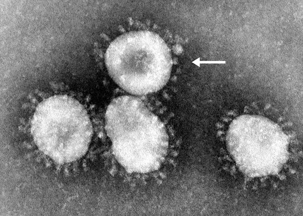
Figure one: Electron micrograph of coronaviruses taken with a transmission electron microscope with the arrow pointing to a single virion. Image taken from Valencia et al. 2020. Original image from Centers for Disease Control and Prevention (CDC)
Much of the research has focused on the spike protein, which gives coronaviruses a “crown-like” appearance on electron micrographs. Scientists at the University of Texas-Austin used cryo-EM to determine the first near atomic-resolution map of the spike protein, which had been locked into a stabilized configuration (Wrapp et al. 2020). A similar effort at the University of Washington used cryo-EM to capture high-resolution views of the spike protein in two distinct conformations (‘open’ vs. ‘closed’) in the same sample, giving structural insights into how the spike protein opens to initiate infection (Walls et al. 2020). A specific region of the spike protein called the receptor-binding domain (RBD) binds tightly to ACE2 receptors on human cells, which allows the virus to gain entry during infection. Several groups have determined high-resolution structures of the RBD bound to the ACE2 receptor using crystallography and cryo-EM to gain insights into this interaction, which is a key target for the development of both vaccines and antiviral therapeutics (Wang et al. 2020, Lan et al. 2020, Shang et al. 2020, Yan et al. 2020). Interestingly, the novel SARS-CoV-2 virus was found to bind to the same neutralizing antibody at the same binding site as the SARS-CoV-1 virus that caused the first SARS epidemic in 2003 (Yuan et al. 2020). This discovery indicates that the spike protein is highly conserved structurally and suggests the possibility of developing antibodies that may broadly neutralize all types of coronaviruses.
Another important target is the main protease (MPro), which yields functional viral proteins translated from viral RNA. Researchers at Shanghi Tech University and the University of Lubeck have solved crystal structures of MPro in complex with two different inhibitors (Jin et al. 2020, Zhang et al. 2020). Recent efforts towards structure-based drug design by researchers in China have allowed them to solve crystal structures of MPro bound to two drug candidates (Dai et al. 2020). Furthermore, a significant endeavor by a team at Diamond Light Source in the UK has provided a high-throughput fragment screening platform to determine crystal structures of MPro in complex with a panel of many different potential inhibitors. Their full 1500-crystal experiment ultimately identified 66 active site fragments.
Finally, the RNA-dependent RNA polymerase is another key target, due to its critical role in copying the RNA viral genome. Researchers in China have used cryo-EM to determine the atomic-level structure of this protein complex alone (Gao et al. 2020), as well as bound to an antiviral drug called Remdesivir (Yin et al. 2020). This drug was originally developed by Gilead Sciences to treat Ebola, but has recently been shown to help some patients recover faster from advanced COVID-19 symptoms in an NIH clinical trial.
While the rapid advancements outlined here are truly remarkable, further technological developments related to crystallography and cryo-EM have the potential to further streamline and expedite the fight against this virus. To aid in this all-important effort, SPT Labtech has been providing next-generation tools for sample preparation, such as the chameleon for cryo-EM grid vitrification and liquid handling robots such as the mosquito and dragonfly to enable efficient crystallographic screening, resulting as seen in recent high-impact publications (Lan et al. 2020, Wang et al. 2020).
For further information, explore how we are supporting our customers through COVID-19.