


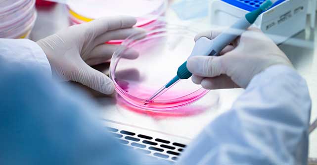
Cell-based assays are experimental methods that use living cells to evaluate biological activity, toxicity, or function. They're essential in drug discovery, toxicology, and biomedical research because they offer human-relevant data and help reduce reliance on animal models.
- Applications
-
Products
-
Liquid Handling
- firefly Accelerate genomic research with innovative all-in-one, compact liquid handling
- mosquito Nanolitre liquid handling technology performs ‘traditional’ tasks at a fraction of the volume, and higher speeds
- dragonfly Delivers accurate and repeatable nanolitre to milliliter dispensing
- apricot Automated liquid handling instrumentation for convenient general use across your entire team
- Sample Preparation
-
Sample Management
- comPOUND A scalable, reliable, and secure compound management solution
- BioMicroLab Easy-to-use sample management automation instruments
- arktic Robust biospecimen storage and management down to -80°C
- lab2lab Novel sample and data transfer network system
- comPACT Reliable and efficient -20°C storage and retrieval has never been more accessible
-
Liquid Handling
-
About
- Company With a focus on liquid handling, sample preparation and sample management, our expert teams create state-of-the-art solutions that scientists and researchers can trust Culture We have one overarching mission: to work together to accelerate life science research. Through our innovative solutions and state-of-the-art tools, we believe we can make a real difference to human health Partners Collaboration is key in our mission to make a real difference to human health. Partnering with application leaders globally, we co-create to solve new challenges across the life sciences. Innovation From the initial prototype through to manufacturing, installation and beyond, we bring a problem-solving mindset and technical expertise to drive innovation
-
Executive Leadership
 Through strategic guidance, visionary thinking, and a relentless pursuit of excellence, our senior executives steer SPT Labtech towards achieving its mission of making a real difference to human health through solving advanced laboratory challenges.
Learn more
Through strategic guidance, visionary thinking, and a relentless pursuit of excellence, our senior executives steer SPT Labtech towards achieving its mission of making a real difference to human health through solving advanced laboratory challenges.
Learn more 
-
View all
Board of Directors
 Our Board of Directors are committed to driving the long-term success and sustainability of SPT Labtech, providing expert guidance and oversight to execute the company’s ambitious commercial strategy.
Learn more
Our Board of Directors are committed to driving the long-term success and sustainability of SPT Labtech, providing expert guidance and oversight to execute the company’s ambitious commercial strategy.
Learn more
-
Knowledge Base
- Resources Our wide range of insightful resources include videos, whitepapers, eBooks, application notes and more Events & Webinars Meet the SPT team at events all over the globe and virtually via our webinars Podcast We chat with innovators and leaders from across the community to gain their unique insights. News Latest news from SPT Labtech globally Blog Our latest blog posts feature trends in research, innovative techniques and new technology
-
08 July, 2026
 Building an Automated State of Mind: How the University of Michigan Advanced Genomics Core Is Rewriting the Rules of Liquid Handling
Continue reading
Building an Automated State of Mind: How the University of Michigan Advanced Genomics Core Is Rewriting the Rules of Liquid Handling
Continue reading
-
01 July, 2026
 Scaling Down & Powering Up: Driving Efficiency in Medicinal Chemistry Through Miniaturization
Continue reading
Scaling Down & Powering Up: Driving Efficiency in Medicinal Chemistry Through Miniaturization
Continue reading
-
13 April, 2026
 SPT Labtech and EMBL GeneCore Collaborate to Advance Fully Walkaway Automation in Genomics Workflows
Continue reading
SPT Labtech and EMBL GeneCore Collaborate to Advance Fully Walkaway Automation in Genomics Workflows
Continue reading
10
- Careers
- Home
- Drug Discovery
- Cell-Based Assays
The Complete Guide to Cell-Based Assays: Exploring the Workflows, Challenges, and Opportunities for Automation
How do scientists study the ways living cells respond to compounds, genetic changes, or their environmental stimuli? One of the most powerful approaches is the use of cell-based assays, which allow researchers to observe cellular behavior in controlled settings. As more labs seek alternatives to animal testing and strive for more predictive, human-relevant data, the role of cell-based assays is rapidly expanding.
But if you ask a group of scientists how they conduct these assays, you're unlikely to hear the same answer twice. That’s because this field is far from standardized. While genomics workflows are often kit-based and protocol-driven, cell-based assays reflect a more experimental, variable, and dynamic approach to lab science.
In this guide, we’ll walk through the key steps involved in cell-based assay workflows from cell culture to cell painting, and highlight the complexity and flexibility researchers must navigate. Along the way, we’ll explore how certain steps can benefit from automation, helping labs reduce manual burden and increase throughput without sacrificing data quality.
What are cell-based assays?
A cell-based assay is any lab technique that uses living cells to study biological processes. These assays are critical in fields like drug discovery, toxicology, immunology, and precision medicine. By observing how cells react to treatments or genetic modifications, scientists can assess viability, function, toxicity, and more, offering insights that test tubes and animal models often can’t provide.
What sets cell-based assays apart is their reliance on living systems. Cells breathe, divide, metabolize, and adapt. This makes them powerful models - but also delicate and difficult to control.
Cell-based assays: A flexible and non-linear workflow
Unlike standardized techniques, there’s no single way to run a cell-based assay. Laboratories mix and match methods based on their needs, scale, cell types, and desired readouts. Some workflows are straightforward, while others involve iterative loops of optimization and retesting.
Still, most assays involve variations of the following methods:
- Cell culture
- 3D cell culture
- Co-culture
- Assay development and media optimization
- Viability testing
- Compound addition
- Toxicity testing
- Cell imaging and staining
- Cell painting
- Transfection
FAQ's
Let’s look at each method in more detail.
Cell culture
Cell culture is the cornerstone of cell-based assay workflows. It refers to the process of growing and maintaining cells in a controlled in-vitro environment, typically within dishes or multi-well plates filled with nutrient-rich media. Researchers often begin with cryopreserved cells stored at -80°C or in liquid nitrogen, and revive them by thawing and transferring them into fresh culture media.Most laboratories rely on 2D culture systems, where adherent cells grow in a monolayer on the surface of a plastic vessel. These vessels range widely in format depending on the experiment's scale, from single petri dishes to 6-, 24-, 96-, 384- and even 1,536-well plates. Once plated, cells metabolize, divide, and release waste into the media. To support healthy cell growth, researchers must carefully monitor pH, temperature, gas composition, and metabolite levels and periodically exchange media.
Cells must also be "split", or passaged, when they reach excessive confluency, meaning they cover the surface of the dish to higher density than optimal for a cell line. Passaging involves detaching the cells from the surface of the plate, mixing them with fresh media, and redistributing them into new vessels to maintain optimal cell density. Scientists may expand cell stocks for future experiments, freeze down excess cells, or prepare cells for downstream assays like drug testing or imaging.
However, cell culture isn't one-size-fits-all. Different cell lines have different requirements, including specific media compositions and gas concentrations. Some cells thrive in basic media formulations, while others need media enriched with multiple supplements, such as serum, growth factors, amino acids, antibiotics, and antimycotics. In cases where cell culture media requirements are unknown, researchers may run optimization experiments with varying formulations.
Full automation of this process is challenging due to the need for integrated incubators, fridges, centrifuges, and complex labware compatibility. While large-scale systems exist, they are often bulky and expensive. Instead, many labs adopt partial automation strategies. For instance, SPT Labtech's solutions can automate certain downstream-compatible steps, such as plating cells from stock vessels into SBS-format plates or exchanging media in higher-density plates.
Our automated systems offer gentle, reproducible liquid handling, making them ideal for preparing cell plates for subsequent assays. Although they do not replace full-scale manual culture, they significantly reduce manual burden for labs working with established cell stocks, especially in high-throughput settings.
By focusing on these compatible phases such as dispensing, plating, and media exchange, researchers can integrate automation where it adds the most value, maintaining flexibility without compromising cell health or assay fidelity.
3D cell culture is a rapidly evolving cell culture approach that is gaining traction as a more physiologically relevant alternative to traditional 2D systems. Unlike monolayer cultures, 3D models allow cells to grow in three dimensions: closely mimicking the architecture, nutrient gradients, and cell-to-cell interactions found in real tissues.
This approach has garnered heightened interest following recent FDA guidance advocating for the reduction of animal testing. The agency's support for "New Approach Methodologies" (NAMs), including 3D culture and organ-on-chip models, reflects a growing recognition that animal models often fail to predict human responses accurately. As such, 3D culture is poised to play a key role in next-generation drug development and toxicity testing.
3D culture systems vary in complexity. Basic models like spheroids consist of a single cell type organized into spherical structures, where cells on the interior and exterior experience distinct oxygen and nutrient conditions. More advanced organoids are self-organizing clusters derived from stem or progenitor cells and include multiple cell types arranged to resemble miniature organs.
To support these structures, researchers commonly use hydrogels - semi-solid matrices that replicate the extracellular environment. These gels provide mechanical support and biochemical cues necessary for tissue-like growth. Matrigel® is a widely used animal-derived example, while synthetic alternatives like GrowDex® and Peptimatrix™ offer reproducibility and reduced biological variability. Each hydrogel comes with its handling challenges, particularly due to viscosity and temperature sensitivity.
Manual pipetting of hydrogels is labor-intensive, challenging to scale up, and prone to variability. Matrigel®, for example, must be kept cold to remain liquid and quickly dispensed, as it will solidify at temperatures above 10°C. These challenges make 3D culture an excellent candidate for automation. SPT Labtech's positive displacement liquid handling solutions like dragonfly®, firefly® and mosquito® have demonstrated effectiveness in dispensing viscous hydrogels with high precision and reproducibility.
Our systems have been validated with both animal-derived and synthetic matrices, supporting 3D formats like domes, embedded cultures, and thicker gel layers across various well-plate formats. Automation makes it possible to scale up these assays enabling higher-throughput formats like 96- and 384-well plates, which are otherwise impractical by hand due to the complexity of hydrogel handling.
By integrating automation into 3D cell culture workflows, researchers can accelerate assay development, perform multi-condition optimization, and reduce variability. As the demand for human-relevant, scalable testing increases, automated 3D culture solutions will be instrumental in driving both scientific and regulatory progress.


High-throughput endometrial organoid plating in Matrigel using SPT Labtech's firefly
Automated high-throughput endometrial organoid plating in 100% Matrigel® using firefly®
How to dispense Matrigel and PeptiMatrix hydrogels using the dragonfly® discovery
Co-culture
Co-culture models aim to capture the biological complexity of real tissues by growing multiple cell types together either in shared environments or separated by permeable barriers that allow chemical signaling. Unlike conventional 2D culture, which typically involves a single cell line, co-culture investigates how different cells interact, communicate, and influence each other’s behavior. These interactions are often chemical in nature, involving the secretion of signaling molecules or metabolic byproducts that affect neighboring cells.
There’s no single way to perform a co-culture. Some researchers simply mix two or more cell lines together in a standard cell culture dish. Others adopt more complex strategies, such as layering different cell types in a 3D matrix or using transwell systems where cells are physically separated but can exchange soluble factors. In 3D co-culture systems, scientists might arrange different cell types in stacked hydrogel layers, or co-embed them within a single matrix to simulate intricate in vivo-like architectures.
This flexibility is part of what makes co-culture both powerful and challenging. Because different cell types often have varying media requirements, optimal seeding densities, and growth rates, a critical part of any co-culture setup is assay optimization. Researchers frequently perform titration experiments such as varying the ratios of two cell types across a plate, to determine the ideal combination for functional readouts.
Automation can be especially helpful in these optimization phases. Products like SPT Labtech’s dragonfly discovery or firefly allow for precise liquid handling and multiplexed dispensing, making it easy to set up ratio gradients or media compatibility tests. This helps reduce manual pipetting burden and ensures more reproducible results.
As co-culture continues to evolve from simple 2D mixes to complex 3D environments, it offers researchers a more accurate lens into tissue-level biology. With automation tools that support flexible formats and complex liquid handling, labs can scale up co-culture experiments while maintaining the precision needed for meaningful interpretation.

Designing the future of cultivated meat: An Uncommon journey to cost-effective stem cell growth media and RNA delivery
GSK collaboration to accelerate life science research
Assay development and media optimization
Assay development and media optimization are foundational steps in designing meaningful cell-based experiments. At its core, an assay is a structured experiment that produces a measurable biological readout: whether that’s cell viability, morphology, or response to a treatment. For example, a simple assay might involve treating cells with a compound and measuring survival using a viability dye or fluorescence signal.
While some assays come in ready-to-use kits with standard protocols and reagents, researchers often find that real-world applications require customization. Different cell types (especially patient-derived or genetically modified lines) may not behave as expected under default conditions. This creates a need for assay optimization, where scientists tweak variables like incubation time, compound concentration, or detection method to ensure reliable results.
Media optimization is a more specific, yet equally critical, subset of this process. Many cell lines depend on customized nutrients and growth factors to thrive. Researchers often start with a base media formulation and add supplements (often called additives) that must be titrated to find the right concentrations. This is particularly important for sensitive or rare cell types that may die under suboptimal conditions. For instance, while a robust cancer cell line may tolerate a wide range of conditions, a delicate patient-derived sample could fail without precise media tuning.
Because of the many variables involved, assay and media optimization can quickly become complex. That’s where tools like design-of-experiment (DoE) software come in - providing statistical frameworks for testing multiple variables efficiently. Automation solutions such as SPT Labtech’s dragonfly discovery and firefly are ideal for these tasks. With support for up to 10 independently controlled syringes, dragonfly discovery enables researchers to dispense precise volumes of multiple reagents across a wide range of conditions in a single run.
The integration with Synthace, a DoE software platform built for biologists, adds even more value. Researchers can design, model, and execute complex optimization studies (from media titration to multi-variable assay configurations) while minimizing manual setup time. This partnership has already been showcased in projects like the British Heart Foundation’s cardiac model development, where researchers used dragonfly to mix different cell types and refine culture conditions for heart tissue modeling.
In essence, assay development and media optimization aren’t just about fine-tuning - they’re about making sure experiments yield data that’s reproducible, relevant, and biologically insightful. With automation, labs can scale up their optimization efforts, reduce human error, and explore more experimental space than manual methods allow.
Viability testing
Viability testing is a core component of many cell-based assays, offering a straightforward readout on one of the most fundamental biological questions: are the cells alive? Scientists use viability assays to assess whether a treatment such as a compound, genetic modification, or environmental condition, has a toxic or tolerable effect on cells. It’s also a common quality control measure during routine cell culture or therapeutic preparation.
There are many ways to measure viability. Some are highly manual, like visually inspecting cells under a microscope. Others rely on specialized instruments, such as flow cytometers or dedicated counters like the NucleoCounter NC-202 from Chemometec. However, these instruments typically fall outside the scope of general-purpose automation solutions due to their proprietary hardware, consumables, and workflows.
For most labs, chemical or reagent-based assays are the most accessible method for measuring viability. These often use dyes or luminescent substrates that distinguish between live and dead cells. A popular example is CellTiter-Glo from Promega, which produces a luminescent signal proportional to the amount of ATP present: an indicator of metabolically active, viable cells.
While many viability assays remain compatible with imaging or slide-based workflows, plate-based formats are especially amenable to automation. As researchers move between formats e.g., from culture plates to slides or flow cytometry vials, it becomes clear that no single system can cover every need. But when viability testing is performed in high-density plate formats using chemical assays, automation delivers significant advantages in throughput, consistency, and ease of use.
Compound addition is a pivotal step in most cell-based assays, where researchers introduce test substances (typically drugs or chemical agents) into cell cultures to observe biological responses. Depending on the experimental aim, these compounds may be the central focus of investigation, as in drug screening or mechanism-of-action studies, or they may act in a supporting role, serving as assay substrates, inhibitors, or readout reagents.
Compounds are usually introduced in liquid form, often diluted in DMSO or aqueous solution. A frequent requirement in these workflows is the preparation of accurate concentration ranges, often through serial dilution, to characterize dose–response relationships or determine potency (e.g., EC₅₀, IC₅₀). Automated instruments enable researchers to generate these dilution series reproducibly, reducing variability associated with manual pipetting and allowing efficient implementation of design-of-experiment approaches.
When working at large scale or with limited compound stocks, liquid handlers capable of nanolitre dispensing such as mosquito enable accurate delivery of concentrated solutions while minimizing reagent waste and exposure to solvents like DMSO. This is particularly relevant in miniaturized or high-content assays, where maintaining consistent ratios of compound to cell culture volume is essential for reproducible results.Toxicity testing
Toxicity testing is a critical outcome-focused application within the cell-based assay workflow. While it isn’t always a standalone step, it overlaps significantly with assay development, compound addition, and viability testing. The goal is simple yet essential: to determine whether a given compound (often a drug candidate) has harmful effects on cells, and at what concentration those effects occur.
In practice, toxicity assays typically involve treating cells with varying doses of a compound and measuring biological responses over time. This may include assessing cell death, proliferation rate, or morphological changes. Many of the same tools used in viability assays like ATP-based luminescent readouts (e.g., CellTiter-Glo) are also used to infer toxicity.
Because toxic responses can depend on both dose and exposure time, these experiments often involve a matrix of conditions. Researchers might explore multiple concentrations across several incubation periods to map the compound’s safety and efficacy window. This makes toxicity testing a natural candidate for automation, particularly when paired with design-of-experiment (DoE) approaches.
Though not labeled as a discrete “step” in linear workflows, toxicity testing is a central aim of many assays and an integrative outcome of several upstream processes. It highlights the non-linear, interconnected nature of cell-based workflows where decisions around compound delivery, incubation timing, and readout formats all converge to determine a compound’s safety profile.
Automation helps streamline this complexity, making it easier for scientists to identify toxic effects early, refine dose-response curves, and inform go/no-go decisions in drug development pipelines.
Cell imaging and staining
Cell imaging enables visualization and interpretation of cellular responses in a wide range of assays, including viability, morphology, and phenotypic screens. It allows scientists to see what’s happening inside cells, such as whether they’re alive or dead, dividing or damaged, or expressing certain markers of interest. Cell staining is a laboratory technique that often precedes cell imaging and uses colored or fluorescent dyes to make cells and their internal structures more visible under a microscope.
Cell staining typically involves adding one or more dyes to cells to highlight specific cell types or sub-cellular structures. Some stains are selective for live cells, others for dead cells, and some can distinguish between them using different fluorescence channels. For example, a common combination might stain live cells green and dead cells red. More advanced protocols may involve multiple dyes to label nuclei, cytoskeletons, mitochondria, or other organelles.
Staining workflows often include several steps: adding the dye(s) to the cells, incubating for a set amount of time, aspirating excess dye, and washing with a buffer to reduce background signal. This washing step is key to generating clear imaging data, as leftover dye can obscure cellular details and compromise data quality.
Staining workflows often include several steps: adding the dye(s) to the cells, incubating for a set amount of time, aspirating excess dye, and washing with a buffer to reduce background signal. This washing step is key to generating clear imaging data, as leftover dye can obscure cellular details and compromise data quality.
Automation can significantly streamline these workflows. For example, SPT Labtech’s products - firefly, dragonfly discovery, and apricot S3 - support gentle and precise liquid handling for stain addition, incubation steps, and complex wash cycles. Instruments with more advanced software capabilities are especially well-suited for multi-step protocols that require precise timing or sequential reagent addition.
Whether using a single stain or conducting multiplexed imaging with several dyes, automation provides consistency and reduces the manual burden of repetitive pipetting. This is particularly beneficial in high-throughput environments, where preparing dozens (or hundreds) of cell imaging plates by hand would be labor-intensive and error-prone.
Ultimately, cell imaging bridges the gap between functional assays and visual interpretation, offering rich, spatially resolved data. Aided by the right automation tools, researchers can scale cell staining and imaging protocols confidently and reliably, supporting everything from simple viability checks to complex phenotyping.
Cell painting
Cell painting is a specialized, high-content imaging technique that builds on traditional cell staining but applies a standardized, multiplexed approach. The goal is to use a defined set of dyes, typically five to seven, that label different cellular compartments such as the nucleus, cytoskeleton, mitochondria, and endoplasmic reticulum. This creates a “morphological fingerprint” that captures the phenotypic state of the cell.
What sets cell painting apart is that it doesn’t require prior knowledge of a compound’s mechanism of action. Instead, researchers infer effects based on deviations from well-established morphological baselines. For example, if a treated cell shows an abnormal nucleus shape or altered mitochondrial structure compared to untreated controls, it suggests that the compound has a biological effect even if the exact molecular target or mechanism of action is unknown.
This approach is particularly powerful when combined with machine learning and reference datasets. The field has robust open-source libraries for healthy and perturbed cell states, enabling researchers to compare their results against large-scale annotated data. Cell painting is increasingly used in drug discovery, phenotypic screening, and toxicology due to its ability to extract rich, unbiased information from a single assay.The protocol typically involves sequential dye addition, timed incubations, and multiple wash steps to remove background signal, all of which can be efficiently automated. SPT Labtech’s dragonfly discovery platform is especially well-suited for this, thanks to its precise control of liquid handling, flexible software, and compatibility with SBS-format plates.
SPT Labtech has also demonstrated strong proof-of-concept work with innovative partners like Semarion, who are using magnetic, barcoded microchips for miniaturized 2D cell culture. These chips support precise multiplexing and simplify imaging workflows. Because the chips can be frozen, thawed, and reused in a standardized manner, they reduce variability and make cell painting more scalable.
Automating cell painting enables high-throughput phenotypic profiling with greater consistency and efficiency. As the field continues to grow with increasing data sharing, community standards, and start-up innovation, solutions that support miniaturization, flexibility, and gentle reagent handling will be key to driving progress.
Transfection
Transfection refers to the process of introducing external materials - typically DNA, RNA, or other nucleic acid constructs - into living cells. These constructs may be used to modify gene expression, introduce reporter genes, or test the cellular response to various genetic edits. Because cellular membranes act as selective barriers, larger molecules like DNA cannot easily enter cells unaided, and dedicated techniques are required to permeabilize the membrane temporarily.
Common methods include electroporation, which uses short electrical pulses to open transient pores in the cell membrane, and chemical or lipid-based reagents that facilitate uptake. While effective, these methods can be harsh and may result in cell death, so optimizing transfection protocols requires careful consideration of conditions such as voltage, construct concentration, and recovery time.
Transfection is not a standalone event but rather part of a broader experimental pipeline. It typically follows upstream steps like cell culture, plating, and assay setup, and is followed by downstream processes such as media exchange, viability assays, and imaging to assess transfection efficiency.
As transfection continues to be a gateway to advanced cell engineering, gene editing, and functional genomics, automation solutions that integrate seamlessly into broader workcells will be key to improving reproducibility and throughput, especially when experiments involve iterative rounds of modification, analysis, and selection.
Flexibility matters: no two cell-based assay workflows are the same
As this guide illustrates, cell-based assays are anything but linear. Labs jump between steps, repeat processes, and use wildly different formats depending on their research goals. This flexibility is part of what makes the field so powerful and so challenging.
That’s why automation solutions need to be equally flexible. Companies like SPT Labtech, which design modular instruments addressing specific bottlenecks, including plating hydrogel-embedded organoids for 3D cell culture, dispensing sensitive reagents, and optimizing complex assays, will be critical to the future of cell-based assay workflows.
Final thoughts: the future of cell-based assays
As regulatory bodies shift away from animal models and demand more human-relevant data, cell-based assays are becoming a cornerstone of modern biomedical research. From growing 3D organoids to testing personalized therapies, these workflows offer the versatility and biological insight needed for the next generation of medical breakthroughs.
By investing in automation tools that are gentle, precise, and adaptable, researchers can unlock new possibilities like accelerating discoveries, reducing variability, and doing more with less.
Whether you're just getting started with cell-based assays or scaling up an advanced screening campaign, understanding the steps (and where technology can help) is key to success. As this guide illustrates, cell-based assays are anything but linear. Labs jump between steps, repeat processes, and use wildly different formats depending on their research goals. This flexibility is part of what makes the field so powerful and so challenging.
That’s why automation solutions need to be equally flexible. Companies like SPT Labtech, which design modular instruments addressing specific bottlenecks, including plating hydrogel-embedded organoids for 3D cell culture, dispensing sensitive reagents, and optimizing complex assays, will be critical to the future of cell-based assay workflows.
In reality, it is not always practicable. While many steps like plating cells, media exchange, staining, and reagent dispensing can be automated, other stages (e.g., cell incubation or electroporation) still rely on specialized equipment or manual handling. Automation solutions work best when integrated into modular workflows.
Automation tools from SPT Labtech are optimized for SBS-format plates (96, 384, 1536 wells). While traditional larger dishes and flasks may not be compatible, our systems are ideal for preparing assay-ready plates and handling media and reagent exchanges in high-throughput formats.
Cell painting is a standardized imaging assay that uses multiple dyes to stain specific cell compartments. Unlike simple staining, it captures comprehensive morphological profiles, enabling phenotypic screening and machine learning-based analysis without needing prior knowledge of a compound’s mechanism.
Automation is especially useful for upstream and downstream processes around transfection such as cell plating, media addition, reagent dispensing, and post-transfection assays. While the transfection step itself (e.g., electroporation) is typically manual or device-specific, automated liquid handling improves consistency and throughput across the rest of the workflow.
Yes. Our platforms are frequently integrated into larger automated workcells alongside robotic arms, incubators, and specialized instruments. This allows labs to automate more of their workflow while maintaining flexibility and precision.