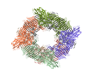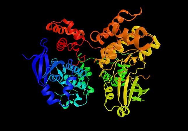


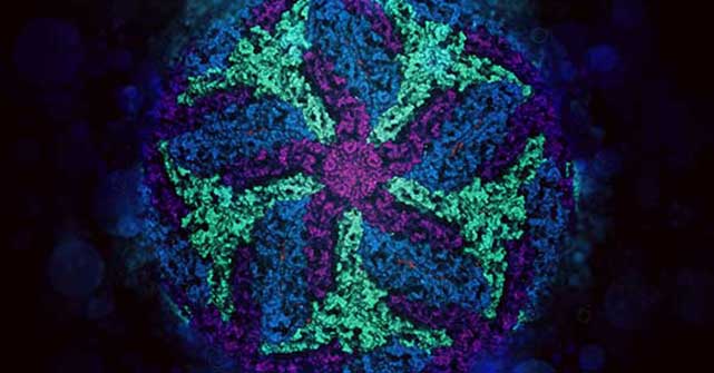
Cryo-EM has ushered in a new era of scientific discovery with the production of increasingly higher-resolution structural information. Advances in hardware and software have now opened up the technique to more widespread adoption in basic research and drug discovery. Cryo-EM is used to visualize a range of biological specimens from large cellular organelles (using cryo tomography) all the way down to the near-atomic resolution of single-particle analysis and micro-electron diffraction.
The role of cryo-EM within ‘integrative’ structural biology
Related Products
chameleon®
chameleon automates the consistent application of samples to high-quality foil grids for cryo-EM analysis, saving time and improving research outcomes across a range of projects
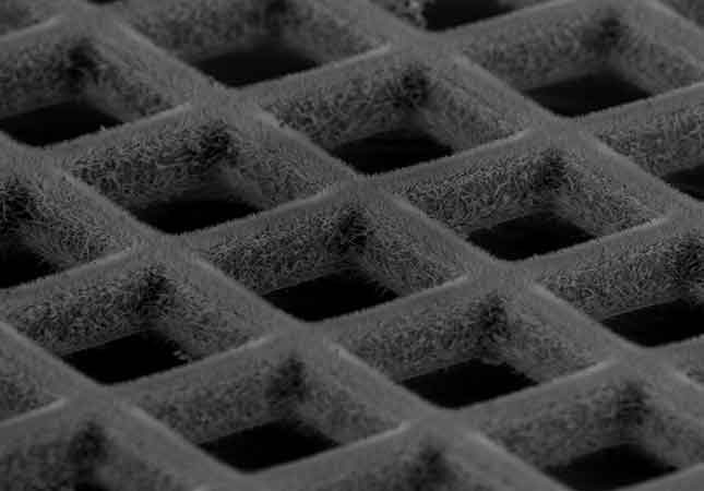
Explore chameleonQuantifoil
Market-leading sample supports enable cryo-EM researchers to optimize data quality.
Explore Quantifoil