


Sample Prep Series Part 1: Preparing your sample for vitrification
28/04/2021
Share this blog
28/04/2021


Sample preservation in vitrified ice (vitrification) is widely acknowledged as the first step in the cryogenic electron microscopy (Cryo-EM) workflow. However, prior to sample preparation researchers must consider the sample quality and determine its suitability for high resolution structure determination. A good understanding of these key considerations along with the right instrumentation is vital to beginning any Cryo-EM project and its subsequent success.
Before pursuing a cryo-EM structure it must be determined that the sample purity is high (>99%, a single band in SDS-PAGE gel), monodisperse in solution and homogeneous in nature (single peak in SEC profile). Particle heterogeneity, both compositional and conformational can lead to difficulty in producing particle image “classes” necessary for building 3D reconstructions from the 2D data. This loss of information limits the quality of the resulting 3D maps and therefore the answerable biological questions.
The biological specimen of interest should remain active in a suitable buffer with optimized conditions. When necessary or after storage, functional assays to determine protein activity and thermostability can be utilized. Vitrification requires low volumes of several microliters and depending on the sample, concentrations from 50nM to 5µM.
With advancements in software and data analysis the single particle workflow can deal with partial heterogeneity and some sample impurity. However, obtaining a solution of isolated homogeneous particles of interest is the best guarantee of a successful high-resolution structure.
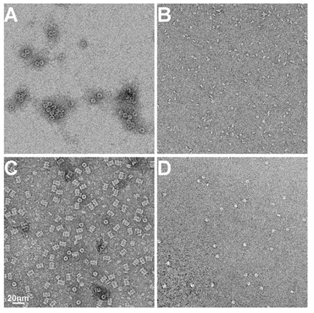 Examples of EM Images of negatively stained protein samples. A) Horse spleen ferritin, B) fragment of antigen binding (Fab), C) archeal 20S proteasome and D) nucleosome. All images at same magnification. The scale bar is 20nm.
It is good practice to assess sample quality and homogeneity prior to vitrification. In addition to biophysical methods like SDS-PAGE, SEC and dynamic light scattering (DLS), the determination of sample homogeneity is routinely accomplished visually using negative stain transmission electron microscopy (nsTEM). This allows the qualitative examination of the particle concentration, composition, and conformational state by fixing the sample solution with stain (e.g., uranyl formate) to a carbon support film on an EM grid. These negatively stained specimens can then be loaded one at a time and examined in simple to use side-entry holder microscopes widely available in EM core laboratories. However, training is required to identify particles of interest and the conditions for “good” and “bad” staining. The time required to perform this analysis is minimal and it provides insight into the specimen quality on the microscopic scale.
The instruments and supplies needed for these methods are readily available in most biochemistry labs and EM core facilities. It is always recommended to optimize sample quality to as high a degree as possible prior to vitrification. Having access to EM specimen prep and analysis nearby to biochemical workflows makes this faster and easier to achieve.

Once biochemical readiness has been determined the sample optimization for plunge freezing can begin. In the next post we will look at the considerations and methods for optimizing samples for conventional sample vitrification for cryo-EM.
D.S. Booth, A. Avila-Sakar, Y. Cheng, Visualizing proteins and macromolecular complexes by negative stain EM: from grid preparation to image acquisition doi: 10.3791/3227 Published: December 22, 2011
B. Carragher, Y. Cheng, A. Frost, R.M. Glaeser, G.C. Lander, E. Nogales, H.-W. Wang, Current outcomes when optimizing ‘standard’ sample preparation for single-particle cryo-EM doi:10.1111/jmi.12834 Published: September 19, 2019
M.A. Cianfrocco, E.H. Kellogg, What could go wrong? A practical guide to single-particle cryo-EM: From biochemistry to atomic models doi: 10.1021/acs.jcim.9b01178 Published: February 20, 2020
R.M. Glaeser, B.-G. Han Opinion: hazards face by macromolecules when confined to thin aqueous films doi: 10.1007/s41048-016-0026-3 Published: July 25, 2017
R.A. Grassucci, D.J. Taylor, J. Frank Preparation of macromolecular complexes for cryo-electron microscopy doi: 10.1038/nprot.2007.452 Published: December 13, 2007
A.J. Noble et. al Routine single particle cryoEM sample and grid characterization by tomography doi: 10.7554/eLife.34257 Published: May 29, 2018
L.A. Passmore, C.J. Russo Specimen preparation for high-resolution cryo-EM doi: 10.1016/bs.mie.2016.04.011 Published: June 16, 2016
G. Skiniotis, D. Southworth, Single-particle cryo-electron microscopy of macromolecular complexes doi: 10.1093/jmicro/dfv366 Published: October 27, 2015
K.A. Taylor, R.M. Glaeser Retrospective on the early development of cryoelectron microscopy of macromolecules and a prospective on opportunities for the future doi: 10.1016/j.jsb.2008.06.004 Published: June 18, 2008